Rose stem, light micrograph

![]()
Wall Art and Photo Gifts from Science Photo Library
Rose stem, light micrograph
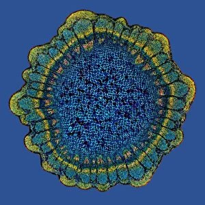
Rose stem. Light micrograph of a cross-section through the stem of a rose (Rosa sp.). The three triangular protrusions are thorns, which protect the plant from being eaten by animals. Each thorn is comprised of the outer epidermis, with a waxy outer cuticle (yellow), a woody layer (brown) and an inner mass of dense parenchyma (pink). The stem is composed of epidermis, bands of parenchyma and chlorenchyma, cortex, vascular bundles - consisting of outer fibres (yellow), phloem (red), cambium (pink) and xylem (brown and pink) - and then the central pith (white). Magnification: x14 when printed 10 centimetres wide
Science Photo Library features Science and Medical images including photos and illustrations
Media ID 6299943
© DR KEITH WHEELER/SCIENCE PHOTO LIBRARY
Cambium Cortex Cross Section Dicot Dicotyledon Internal Microscopy Phloem Pith Plant Anatomy Rose Slice Sliced Stem Thorn Thorns Thorny Tissue Vascular Bundle Xylem Cells Light Micrograph Light Microscope Sectioned
EDITORS COMMENTS
This print showcases the intricate beauty and complexity of a rose stem when viewed under a light microscope. The cross-section reveals the various layers and structures that make up this vital part of the plant's anatomy. The focal point of the image is undoubtedly the three triangular thorns, serving as nature's defense mechanism against potential predators. Each thorn consists of multiple layers, starting with the outer epidermis protected by a waxy cuticle in vibrant yellow. Beneath lies a woody layer in rich brown, followed by an inner mass of dense parenchyma displaying shades of pink. Moving towards the center, we encounter an array of tissues composing the stem itself. These include bands of parenchyma and chlorenchyma responsible for storing nutrients and conducting photosynthesis respectively. Surrounding them is the cortex, providing additional support to this delicate structure. Further inside are vascular bundles essential for transporting water, minerals, and sugars throughout the plant. These bundles consist of outer fibres depicted in yellow, phloem represented by striking red hues, cambium exhibiting soft pink tones, and xylem displaying both brown and pink shades. Finally, at its core lies the central pith portrayed in pure white. With a magnification factor enabling us to appreciate these details when printed 10 centimeters wide at 14 times their original size, this print from Science Photo Library offers viewers an awe-inspiring glimpse into nature's remarkable design found within every rose stem.
MADE IN THE USA
Safe Shipping with 30 Day Money Back Guarantee
FREE PERSONALISATION*
We are proud to offer a range of customisation features including Personalised Captions, Color Filters and Picture Zoom Tools
SECURE PAYMENTS
We happily accept a wide range of payment options so you can pay for the things you need in the way that is most convenient for you
* Options may vary by product and licensing agreement. Zoomed Pictures can be adjusted in the Cart.