Acute erythroid leukaemia, micrograph

![]()
Wall Art and Photo Gifts from Science Photo Library
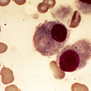
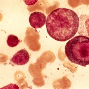
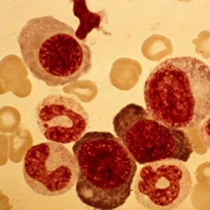
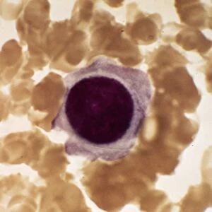
Acute erythroid leukaemia, micrograph
Acute erythroid leukaemia. Light micrograph of blood cells from bone marrow in a case of acute erythroid leukaemia. The cells include dystrophic (degenerated) erythroblasts (nuclei stained dark red), one of which is a megaloblast (centre, enlarged erythroblast) with threefold nuclei. Erythroblasts are the immediate precursors to mature red blood cells as they develop from stem cells in the bone marrow. Acute erythroid leukaemia is a rare form of acute myeloid leukaemia (a blood cell cancer) where the excess blood cell production is of erythroblasts. The pale orange cells are normal red blood cells
Science Photo Library features Science and Medical images including photos and illustrations
Media ID 9241601
© PR. J. BERNARD/CNRI/SCIENCE PHOTO LIBRARY
Acute Myeloid Leukaemia Blood Bone Marrow Cancer Cancerous Cell Biology Cellular Enlarged Erythroblast Haematological Haematology Histological Histopathological Histopathology Leukemia Malignant Oncological Oncology Pathological Pathology Red Blood Cell Abnormal Cells Circulation Circulatory System Condition Disorder Light Micrograph Light Microscope Unhealthy
EDITORS COMMENTS
This print showcases a micrograph of acute erythroid leukaemia, offering a glimpse into the intricate world of blood cells. Taken from bone marrow, the image reveals a multitude of cells, including dystrophic erythroblasts with dark red nuclei. Among them stands out a megaloblast - an enlarged erythroblast with threefold nuclei - emphasizing the abnormality present in this condition. Acute erythroid leukaemia is an uncommon form of acute myeloid leukaemia characterized by an excessive production of erythroblasts. In contrast to the pale orange normal red blood cells seen in the image, these malignant cells represent an unhealthy and disordered state within the circulatory system. The significance lies in understanding that erythroblasts are immediate precursors to mature red blood cells as they develop from stem cells residing in our bone marrow. By studying such histological details through light microscopy, medical professionals gain valuable insights into haematology and pathology. This print not only serves as a visual representation but also highlights the intersection between biology and medicine. It sheds light on oncology and cellular abnormalities within our own bodies while reminding us of both the complexity and fragility inherent in human health. Captured by Science Photo Library, this image offers researchers, educators, and medical practitioners alike an invaluable resource for further study and exploration into conditions like acute erythroid leukaemia.
MADE IN THE USA
Safe Shipping with 30 Day Money Back Guarantee
FREE PERSONALISATION*
We are proud to offer a range of customisation features including Personalised Captions, Color Filters and Picture Zoom Tools
SECURE PAYMENTS
We happily accept a wide range of payment options so you can pay for the things you need in the way that is most convenient for you
* Options may vary by product and licensing agreement. Zoomed Pictures can be adjusted in the Cart.