Sperm fertilising an egg, artwork

![]()
Wall Art and Photo Gifts from Science Photo Library
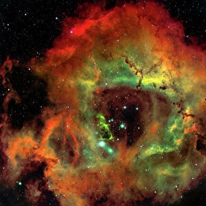
Sperm fertilising an egg, artwork
Sperm fertilising an egg. Cutaway artwork of a human sperm cell (spermatozoon) penetrating an egg cells (ovum) thick outer layer (zona pellucida). The egg cells membrane is at top right. Once inside the ovum, the sperm cell will release the genetic material (deoxyribonucleic acid, DNA) in its head (acrosome) to mix with the DNA in the ovum. The diagram here shows: mitochondria (at base of tail, blue), the protein K81 (base of head, light blue), DNA-containing chromosomes with centromeres (blue with green spheres), m-RNA (messenger ribonucleic acid, yellow), and the proteins lzumo (red) and PLC-zeta (pink)
Science Photo Library features Science and Medical images including photos and illustrations
Media ID 6423072
© HENNING DALHOFF / SCIENCE PHOTO LIBRARY
Acrosome Cell Membrane Centromere Centromeres Chromosome Chromosomes Cut Away Egg Cell Entering Fertilisation Fertilising Fertilization Messenger Ribonucleic Acid Mitochondria Mitochondrion Mrna Ovum Penetrating Proteins Re Production Ribonucleic Acid Sperm Cell Spermatozoon Tail Zona Pellucida Bio Chemistry Biochemical Deoxyribonucleic Acid Protein
EDITORS COMMENTS
This artwork captures the intricate process of fertilization, showcasing the moment when a human sperm cell penetrates an egg's thick outer layer. The cutaway illustration provides a detailed view of this remarkable biological event, highlighting various components involved in the fusion of genetic material. At the top right, we observe the delicate membrane of the egg cell, awaiting its destined partner. As the spermatozoon enters through the zona pellucida, it carries within its head a treasure trove of deoxyribonucleic acid (DNA). This genetic material will soon mingle with that of the ovum to create new life. The diagram further reveals essential elements such as mitochondria located at the base of the tail and protein K81 at the base of its head. Chromosomes containing DNA are depicted in blue with green spheres representing centromeres. Messenger ribonucleic acid (m-RNA) is represented by vibrant yellow hues while proteins lzumo and PLC-zeta add splashes of red and pink respectively. Through this stunning portrayal, Science Photo Library offers us an opportunity to marvel at nature's miraculous processes on a microscopic level. It reminds us that life begins with these intricate interactions between cells and their genetic materials—a testament to both our shared humanity and our unique individuality.
MADE IN THE USA
Safe Shipping with 30 Day Money Back Guarantee
FREE PERSONALISATION*
We are proud to offer a range of customisation features including Personalised Captions, Color Filters and Picture Zoom Tools
SECURE PAYMENTS
We happily accept a wide range of payment options so you can pay for the things you need in the way that is most convenient for you
* Options may vary by product and licensing agreement. Zoomed Pictures can be adjusted in the Cart.