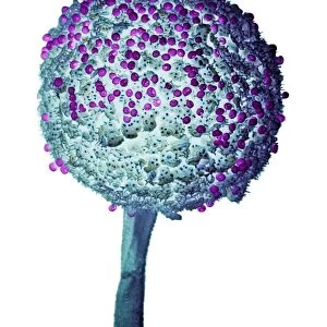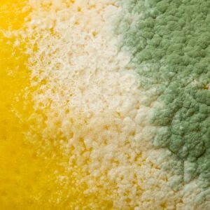
Bread mould, SEM

![]()
Wall Art and Photo Gifts from Science Photo Library
Bread mould, SEM
Bread mould. Coloured scanning electron micrograph (SEM) of fungal hyphae (thread-like structures) growing on the surface of bread (red). These have grown from fungal spores (small white dots) that landed on the bread after drifting through the air. The bread is being digested by the fungi, and this network (mycelium) of hyphae forms. The fungi are also producing new spores. Two types of fungi are seen here. Penicillium sp. is dominant, forming spores from specialised hyphae (conidiophores, feathery green structures, some at lower right). The other fungus is Mucor mucedo, producing spores from its sac-like bodies called sporangia (globular, one at upper right)
Science Photo Library features Science and Medical images including photos and illustrations
Media ID 6292767
© DR JEREMY BURGESS/SCIENCE PHOTO LIBRARY
Bread Bread Mould Conidiophore Conidiophores Digesting Eumycota Food Spoilage Fungal Fungi Fungus Hyphae Mold Mould Mouldy Mycelium Mycology Penicillium Re Production Reproductive Sporangia Sporangium Spore Spores False Coloured Micro Biology Microbiological Spoiling
MADE IN THE USA
Safe Shipping with 30 Day Money Back Guarantee
FREE PERSONALISATION*
We are proud to offer a range of customisation features including Personalised Captions, Color Filters and Picture Zoom Tools
SECURE PAYMENTS
We happily accept a wide range of payment options so you can pay for the things you need in the way that is most convenient for you
* Options may vary by product and licensing agreement. Zoomed Pictures can be adjusted in the Cart.