Arm nerves

![]()
Wall Art and Photo Gifts from Science Photo Library
Arm nerves
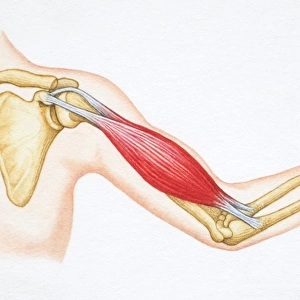
Arm nerves. Historical anatomical artwork of the nerves (white) of the front (palm side) of a human forearm. At left, the superficial (surface) nerves are shown, while at right the deep nerves and muscles (red) are seen. The main deep nerve (down the centre of the arm) is the median nerve. To the right of that is the ulnar nerve. The brachial artery (red) is also seen, dividing into the radial and ulnar branches. The tendons of the hand (grey) are also seen. The superficial arm nerves are the internal and external cutaneous (skin) nerves. Artwork from The Nerves of the Human Body (Ed. Jones Quain, London, 1839)
Science Photo Library features Science and Medical images including photos and illustrations
Media ID 6419566
© SHEILA TERRY/SCIENCE PHOTO LIBRARY
1839 Book Bottom Brachial Artery Connective Tissue Cutaneous Deep Dissection Drawing Fascia Hand Jones Quain Muscles Nerve Nerves Peripheral Radial Skin Superficial Surface Tendon Tendons Text Book Under Side Wrist Median Nervous System Neurological Neurology Section Ulnar
EDITORS COMMENTS
This historical anatomical artwork showcases the intricate network of arm nerves, providing a fascinating glimpse into the inner workings of the human forearm. The print, dating back to 1839, presents a detailed illustration of both the superficial and deep nerves that traverse this vital limb. On the left side of the image, we can observe the surface nerves in white, while on the right side, an array of red lines represents deeper nerves and muscles. Prominently featured is the median nerve running down the center of the arm, accompanied by its neighboring ulnar nerve. Additionally, we can discern the brachial artery dividing into radial and ulnar branches. The grey tendons of hand musculature are also visible in this remarkable piece. Notably depicted are two superficial arm nerves: internal and external cutaneous (skin) nerves. This artwork stems from "The Nerves of Human Body" edited by Jones Quain in London during that era. Transporting us back to a time when medical understanding was rapidly advancing, this illustration serves as a testament to our ongoing exploration and comprehension of human anatomy. It offers invaluable insights into neurology and peripheral nervous system functioning while highlighting connective tissue structures such as fascia. A true treasure for enthusiasts in medicine or history alike, this print from Science Photo Library beautifully captures an important chapter in our scientific journey towards unraveling one's physical composition.
MADE IN THE USA
Safe Shipping with 30 Day Money Back Guarantee
FREE PERSONALISATION*
We are proud to offer a range of customisation features including Personalised Captions, Color Filters and Picture Zoom Tools
SECURE PAYMENTS
We happily accept a wide range of payment options so you can pay for the things you need in the way that is most convenient for you
* Options may vary by product and licensing agreement. Zoomed Pictures can be adjusted in the Cart.